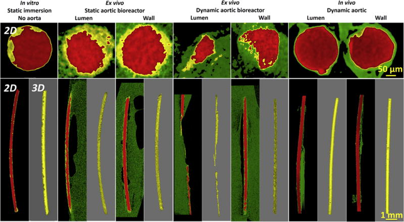
Fig. 4.

Reconstructions of X-ray micro-CT 3-D with representative 2-D slices of Mg wires under static incubation in vitro without aorta, static and dynamic incubation ex vivo in aortic lumen and wall in the bioreactors, and dynamic incubation in aortic lumen and wall in vivo after 3 days. The red parts represent Mg residual, the yellow parts represent degradation products, and the green parts represent host tissue. Note: There are no images for the dynamic ex vivo aortic lumen in the bioreactor, since no residual implant was left. (For interpretation of the references to colour in this figure legend, the reader is referred to the web version of this article.)
