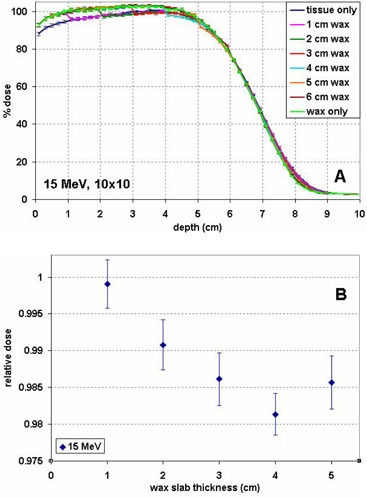
Figure 3.

Percentage depth dose (PDD) curves for a 15 MeV beam (A), normalized to the maximum dose in tissue, for wax slabs of various thicknesses on top of tissue and in tissue or wax only. Relative dose in tissue at the wax–tissue interface (B) for wax slabs of various thicknesses, normalized to the dose at the corresponding depth in a homogeneous tissue phantom.
