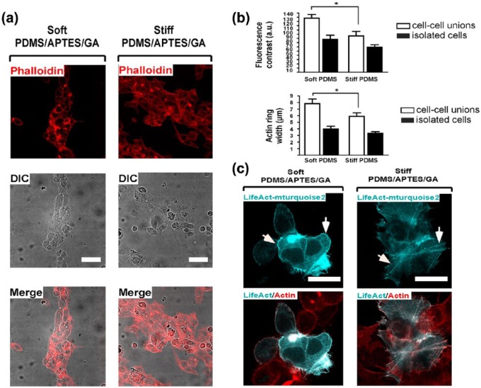
Figure 4.
HepG2 cells confined on soft-PDMS/APTES/GA rather than on stiff scaffolds patterned with COL I in lines exhibit a greater cortical actin arrangement. (a) Actin filaments were observed with Alexa594-coupled phalloidin in HepG2 cells. Cells were seeded at high density on stiff- and soft-PDMS/APTES scaffolds patterned by microcontact-printing with type I collagen in micropatterns. Differential interference contrast (DIC) and confocal microscopy images were acquired 48 h after cell culture. Nuclei were stained with DAPI. Scale bars = 50 μm. (b) Relative F-actin levels were measured as fluorescence contrast (a.u. = arbitrary units) and cortical actin ring width (μm). Data are represented as mean ± SEM (n = 6). *p < 0.021 (cell–cell unions, contrast) and **p < 0.008 (cell–cell unions, width). (c) HepG2 cells were seeded at low density (1 × 10⁵ cells per well) for 24 h on stiff- and soft-PDMS/APTES scaffolds coated with 0.1 mg/mL COL I. F-actin was stained with Alexa594-coupled phalloidin (red) and LifeAct-mTurquoise2 (cyan). Scale bars = 30 μm.

