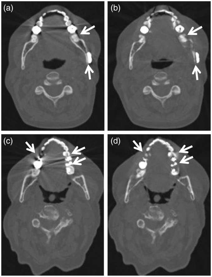
Fig. 3.
Artifactual defect introduction in IMAR images reconstructed with bone window/level settings. wFBP (left column, a, c) and IMAR (right column, b, d) images from two different patients: 53-year-old woman with dental fillings and mandibular hardware (a, b) and 69-year-old woman with dental fillings (c, d). The IMAR images (b, d) introduce artifactual defects along teeth and adjacent bone compared to the wFBP images (a, c) (arrows, a–d). These defects can be seen adjacent to both teeth (b, d) and mandibular hardware (posterior arrow in (b)).

