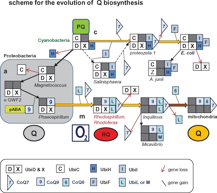
Fig. 3.
—Scheme for the possible evolution of the biosynthetic pathways of Q. The flow scheme illustrates the hypothetical evolution of the pathways of Q biosynthesis presented in this work: a (in gray box on the left), c (top), and m (bottom), all originating from the ancestral pathway of PQ biosynthesis in cyanobacteria (Pfaff et al. 2014). The scheme includes only the enzymes for Q biosynthesis that vary in presence and combination within the genome of proteobacterial taxa; their symbols are presented in the boxed caption at the bottom of the illustration. UbiA is implied to be present in all combinations for its crucial function (see fig. 1 and table 1), whereas UbiB and UbiJ are not considered because their role in Q biosynthesis is not essential and partially unknown (Nowicka and Kruk 2010; Aussel et al. 2014). Gene loss and acquisition is represented by a red thin arrow and thin black line, respectively. A dark gray square indicates the absence of a gene in the combination of enzymes deduced from genomic information. The dashed arrows in the middle indicate a minor alternative route (or intermediate) in pathway c that seems to be present only in deep branching gamma proteobacteria such as Salinisphaera (Pelosi et al. 2016). A genus or species characteristic for each combination of Q biosynthesis enzymes is indicated at the bottom of each set of symbols (cf. tables 1 and 2). Acquisition of other CoQ genes specific to mitochondria (Kawamukai 2015) is not represented at the end of pathway m, bottom right of the scheme. Q in gray circle indicates anaerobically synthesized ubiquinone (pathway a, within the gray square), whereas aerobically synthesized Q is represented as in figure 1. RQ in red circle indicates the Q derivative rhodoquinone that is found in Rhodosprillum and very few other proteobacteria such as Rhodoferax, as well as in anaerobically adapted mitochondria of invertebrates (Hiraishi and Hoshino 1984; Nowicka and Kruk 2010; Müller et al. 2012). α GWF2, alphaproteobacterium GWF2_58_20 (Anantharaman et al. 2016).

