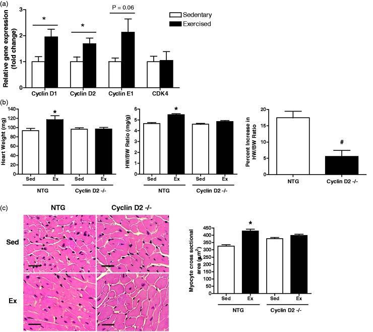
Figure 4.
Expression analysis of a subset of cell cycle genes in sedentary and exercised NTG female mice, and morphometric data, cardiac adaptation, and histological analysis in response to 21 days of exercise for cyclin D2−/− and NTG female mice. (a) Expression of cell cycle genes including cyclin D1, cyclin D2, cyclin E1, and CDK4 in sedentary and exercised (21 days) NTG mice (CB6F1 hybrid line). (b) Morphometric data and cardiac adaptation in cyclin D2−/− and NTG littermate controls: Left: heart weight. Middle: heart weight normalized for body weight (HW/BW) ratio. Right: percent increase in HW/BW ratio. (c) Cardiomyocyte cell size analysis: Left: left ventricular cross-sections stained with hematoxylin and eosin (scale bar = 25 µm). Right: quantification of myocyte cross sectional area (n = 3–4 per group, 3–4 regions per heart, 5–10 cells per region). *, P < 0.05 relative to sedentary counterparts. n = 5–14 animals per group. Data are reported as mean ± SE. (A color version of this figure is available in the online journal.)

