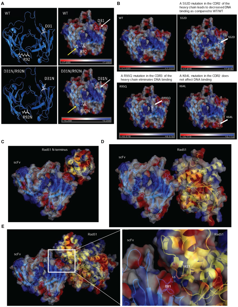
Figure 7.
A molecular model of the 3E10 scFv supports experimental evidence of the DNA binding pocket and the interaction with human RAD51. (A) An electrostatic map generated from a predicted structure of the 3E10 scFv. The D31N mutation is marked with white arrows, while the R92N mutation is marked by yellow arrows. (B) Additional point mutations that have been previously published as having an effect on 3E10 scFv's DNA binding ability were plotted on the generated predicted structure of the 3E10 scFv. (C) A potential model of the interaction between the N-terminus of RAD51 and the 3E10 scFv. (D) A potential model of the interaction between the human RAD51-BRCA2 BRC repeat complex (PDB 1N0W) and the 3E10 scFv. (E) A potential model of the interaction between the Saccharomyces cerevisiae RAD51 structure (PDB 3LDA) and the 3E10 scFv. RAD51 is represented by the yellow ribbon structure in C–E.

