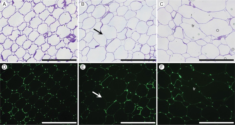
Fig. 7.
Immunofluorescence labelling of cross-sections of the cortex tissue of root segments S1 (A, D), S3 (B, E) and S5 (C, F) using de-esterified backbone-directed antibody (CCRC-M38). Labelling is more prominent in cell intersections (triangular junctions) in S1 and decreases towards S5, but can still be seen in cell walls (tr, trabeculae) in regions surrounding aerenchyma. Scale bar = 100 µm.

