Fig. 8.
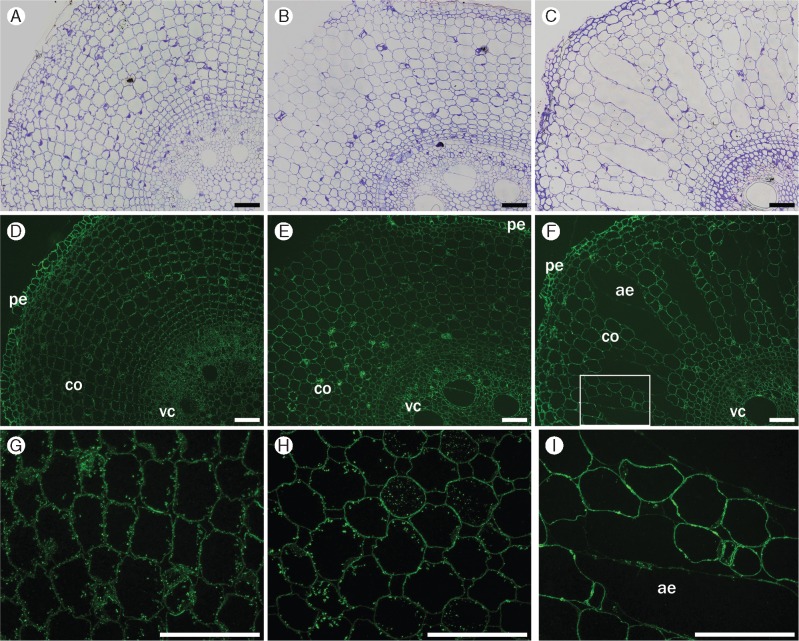
Immunofluorescence labelling of transverse cross-sections from root segments S1 (A, D, G), S3 (B, E, H) and S5 (C, F, I) using the pectic arabinogalactan-directed antibody CCRC-M80. This antibody labels all cell walls in all segments (pe, peripheral tissues; co, cortex; vc, vascular cylinder) (D–F), except for those forming trabeculae inside the aerenchyma (ae). In S1, this antibody also shows labelling of cytoplasmic content closer to inner-facing walls in the cortex (G); labelling is weaker in S3 (H) and absent in S5 (I). The rectangle in (F) is an enlargement of the region shown in I. Controls (A–C) were stained with toluidine blue. Scale bar = 100 µm.

