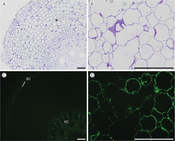
Fig. 9.
Immunofluorescence labelling of cross-sections of root segments using arabinogalactan-directed antibodies JIM14 (S5; A, C) and JIM19 (S1; B, D). (C) the speckled binding pattern of JIM14 monoclonal antibody, labelling cytoplasmic contents of cells from the sclerenchymatous cylinder (sc) and vascular cylinder (vc) in S1. (D) JIM19 binds to cytoplasmic material and cell walls of S5, but binding weaker to walls surrounding the aerenchyma. Controls (A, B) were stained with toluidine blue. Scale bar = 100 µm.

