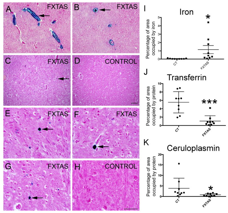
Figure 1.

Iron bound to hemosiderin (blue) accumulated in the putamen in FXTAS. Iron (blue) in sagittal (A) and coronal (B) sections of capillaries showed a great amount of iron in the walls and within the capillaries. Iron was present in the putamen parenchyma in FXTAS (C, E-G) but not in control tissue (D, H). Arrows point to iron deposits. Tissue is co-stained with nuclear fast red. I-K: The amount of iron was increased while the amount of Tf and Cp was decreased in the putamen in FXTAS. Iron: blue, Perl’s method; Tf and Cp: brown, immunostaining. Tf: Transferrin; Cp: ceruloplasmin. Scale bar: A-D: 200 μm; E-H: 100 μm.
