Figure 3.
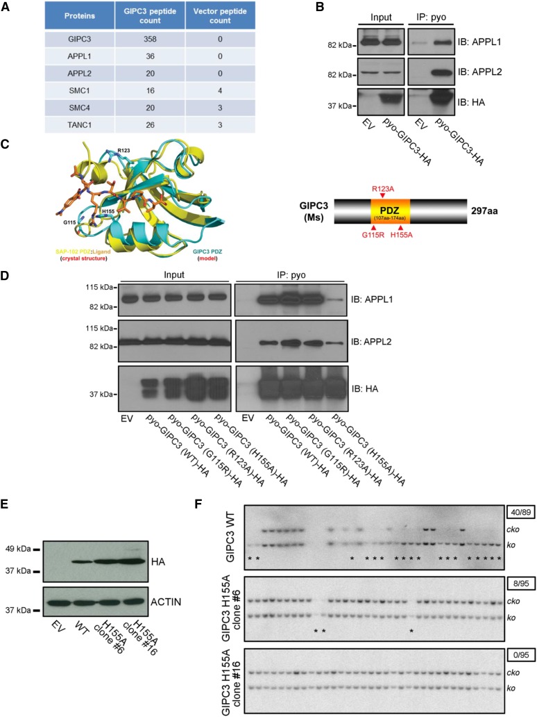
APPL1/2 contributes to the rescue effect of GIPC3 overexpression (A) List of top candidates of GIPC3 interaction proteins as revealed by IP-MS. (B) Western blot of immunoprecipitation in HEK293T cells showing interaction of GIPC3 with APPL1 and APPL2. GIPC3 with Pyo and HA tag (pyo-GIPC3-HA) were ectopically expressed in HEK293T cells. EV, empty vector. (C) Left, schematic illustration showing the superposition of the model of the GIPC3 PDZ domain with the crystal structure of the SAP-102 PDZ domain in complex with a fluorogenic peptide-based ligand (SAP-102:Ligand, PDB entry 3JXT). The polypeptide chains are shown as ribbon diagrams with helices as spirals, strands as arrows, and loops as tubes. The ligand and side chains are shown as stick models. Color schemes are shown and the three predicted residues are labeled. Right, schematic diagram of domain structure of mouse GIPC3 protein and point mutations in the PDZ domain that were generated. (D) Western blot of immunoprecipitation in HEK293T cells showing differential interaction of GIPC3 wildtype (WT) and mutated GIPC3 (G115R, R123A, and H155A) with APPL1 and APPL2. GIPC3 WT and mutants with Pyo and HA tag were ectopically expressed in HEK293T cells. (E) Western blot showing expression of HA tagged GIPC3 in GIPC3 WT and H155A (#6 and #16) stable expressing mESC clones. (F) Southern blot showing differential rescue efficiency of Brca2ko/ko mESC lethality in GIPC3 WT and H155A stable expressing mESC clones (#6 and #16). Asterisks point the rescued Brca2ko/ko clones. Numbers in the upper right boxes indicate rescued clone number over total examined clone number.

