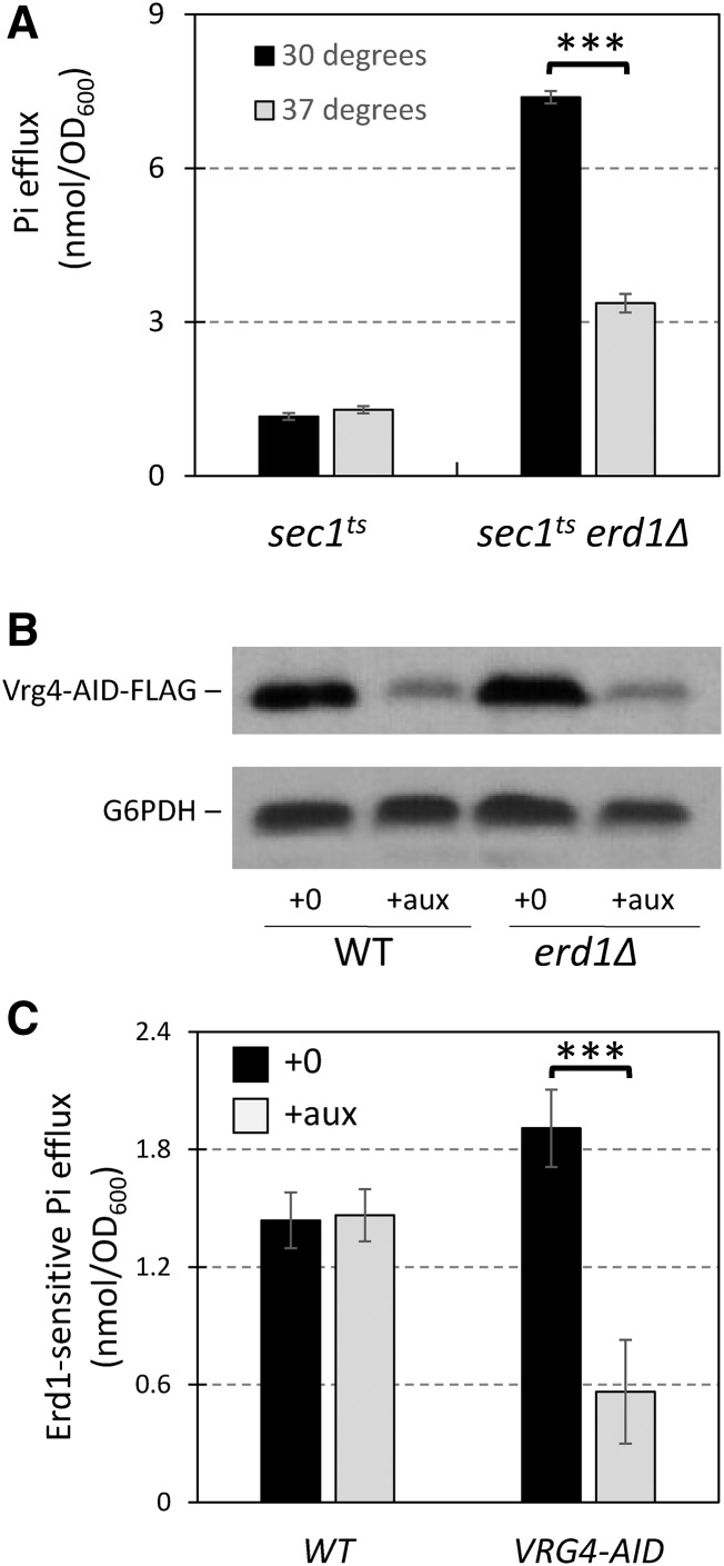
Figure 7.
Erd1-sensitive losses of Pi depend on exocytosis and on transport of GDP-mannose into the Golgi complex. (A) Pi losses were measured after 2 hr incubation as in Figure 6A except that sec1-1ts and sec1-1ts erd1∆ mutant strains were used at a permissive temperature (30°; black bars) or a non-permissive temperature (37°; gray bars) during the incubation in Pi-free medium. (B) Western blot of Vrg4-AID-FLAG strains after 1 hr exposure to 100 µM auxin in SC medium. (C) Erd1-sensitive losses of Pi (i.e., the difference between erd1∆ and ERD1 pairs of strains) were measured for wild-type and Vrg4-AID-FLAG strain backgrounds that were exposed to 100 µM auxin (gray bars) or not (black bars) starting 1 hr before the shifts to Pi-free medium. *** P < 0.001.

