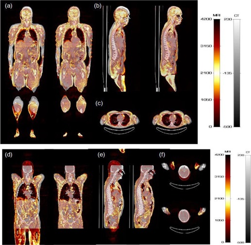
Figure 1.

Representative clinical studies of two patients showing 3D fused MRI and CT images before and after image registration: (a) and (d) coronal, (b) and (e) sagittal, and (c) and (f) transaxial views. For each image pair, left and right panels represent the same image before and after registration, respectively.
