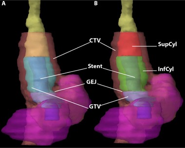
Figure 1.

Coronal volume 3D reconstruction (a) showing the esophagus (yellow), with a self‐expanding stent (SES blue) extending past the gastroesophageal junction (GEJ) into the stomach (magenta) with the GTV (cyan) and CTV (pink) visible; Coronal volume 3D reconstruction (b) now showing post‐stent SupCyl and InfCyl structures (red and green, respectively). When fusing the pre‐stent to the post‐stent CT scans, priority was given to overlaying the SupCyl and InfCyl volumes of each scan, especially in the craniocaudal direction.
