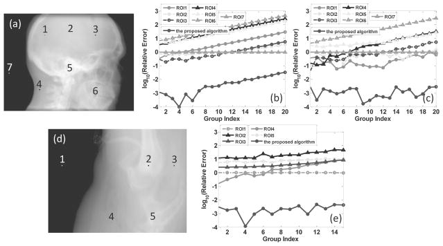
Figure 5.
Comparison results between the proposed algorithm and the homogeneous area based algorithm. The first and the second rows correspond to the head-neck case and the prostate case, respectively. Sub-figures (a) and (d) are projection images in a certain view, where seven and five selected areas are annotated by the numbers, sub-figures (b), (c) and (d) are the log scaled relative errors of the estimated noise levels with both the homogeneous area based algorithm and the proposed algorithm, corresponding to the cases of analytical, MC and the spotlight-mode simulation experiments, respectively.

