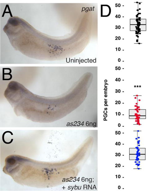
Fig. 3.

Reduction of PGCs in sybu-depleted embryos. (A–C) Whole-mount in situ hybridization for pgat mRNA. (A) control uninjected embryo, (B) sybu-depleted embryo, (C) depleted embryo rescued with injected sybu mRNA (5 pg). (D) Scatter plots of PGC numbers in experimental groups corresponding to the adjacent panel. Dots represent individual in which PGCs were counted (sum of three separate host-transfer experiments); dark line represents the median; shaded areas correspond to the limits of the first and third quartiles (25th/75th percentiles) and whiskers (dotted line) extends out to 1.5× the interquartile range. P < 0.001 by Kruskal-Wallis test; *** = P < 1.0e-11 by Dunn’s post hoc test w/2-sided Bonferroni adjustment.
