
Figure 5.
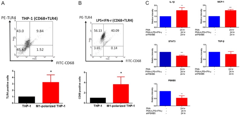
PMA, LPS and IFN-γ treatment promoted THP-1 cells differentiation to M1 macrophages. A, B. The effect of PMA, LPS and IFN-γ treatment on the number of M1 macrophages. THP-1 monocytes were treated with PMA (320 nM) for 6 hours and then cultured with PMA plus LPS (100 ng/ml) and IFN-γ (20 ng/ml) after 24 hours. TLR4 (M1 macrophage marker)-positive and CD68 (macrophage differentiated marker) positive fraction were analyzed using flow cytometry in THP-1 monocytes and M1-polarized THP-1 macrophages. Values are the average of assays performed in triplicate. Error bars represent SD (n=3), P value < 0.05 consider significantly. C. M1 Macrophage markers is expressed in PMA-treated THP-1 macrophage. MCP-1, IL-1β (a marker for M1 macrophage) and TGF-β (a marker for M2 macrophage) mRNA were measured in THP-1 cells and M1-polarized THP-1 macrophages. THP-1 monocytes were treated with PMA (320 nM) for 6 hours and then cultured with PMA plus LPS (100 ng/ml) and IFN-γ (20 ng/ml) after 24 hours. The mRNA levels were measured by RT-PCR and data were normalized according to GAPDH mRNA level and presented as a value relative to that for undifferentiated THP-1 monocytes. Values are the average of assays performed in triplicate. Error bars represent SD (n=3). *p < 0.05.