
Figure 1.
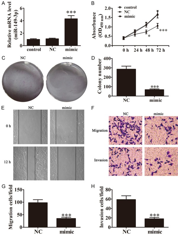
Expression of miR-149-3p inhibits the proliferation, invasion, and migration of BCa cells. A. Expression of miR-149-3p in UM-UC-3 cells was measured by Rt-PCR after transfection with miR-149-3p mimics for 48 h (n=3). Data are presented as the mean ± SD (***P < 0.001 versus control group). B. The CCK-8 assay showed that the relative viability of UM-UC-3 cells treated with miR-149-3p mimics was significantly lower than NC-treated cells (n=5). Data are presented as the mean ± SD (*P < 0.05, ***P < 0.001 versus control group). C and D. Colony formation assay show that the colony formation rates of miR-149-3p mimic-transfected UM-UC-3 cells were lower in contrast with NC-transfected cells (n=5). Data are presented as the mean ± SD (***P < 0.001 versus NC group). E. Wound healing assays indicated that miR-149-3p over0expression reverses the migration of UM-UC-3 cells. F-H. Transwell assays confirmed that miR-149-3p over-expression inhibited migration and invasion of UM-UC-3 cells (n=5). Data are presented as the mean ± SD (***P < 0.001 versus NC group).