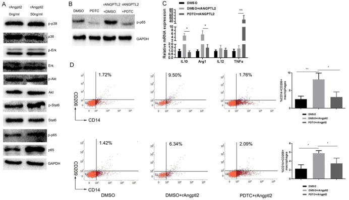
Figure 3.
Angptl2 promoted M2 macrophage polarization via the p65 NF-κB signaling pathway. A. Western blot of phospho-p38 MAPK, p38 MAPK, phospho-Erk, Erk, phospho-Akt, Akt, phospho-Stat6, Stat6, phospho-p65, p65 NF-κB and GAPDH in THP-1-derived macrophages with or without the addition of rAngptl2 (50 ng/ml, 24 hours). B. Western blot of p-p65 NF-κB in THP-1-derived cells with or without pretreatment of PDTC (100 μM in DMSO for 30 min) before the addition of rAngptl2. C. RT-PCR analysis of M1/M2 markers in THP-1-derived cells with or without pretreatment of PDTC before the addition of rAngptl2. D. Flow cytometry analysis of THP-1-derived cells with or without PDTC pretreatment before the addition of rAngptl2. The percentage of CD14+/CD206+ and CD14+/CD209+ cells and quantitative data are shown. *p<0.05, **p<0.01.

