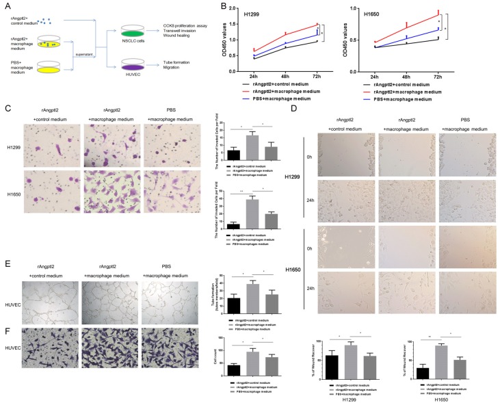
Figure 4.
Angptl2-promoted macrophages enhanced the proliferation, invasion and migration of NSCLC cells and the tube formation and migration of HUVECs in vitro. A. A schematic representation of the experimental approach used in this part. B. NSCLC cells were incubated with the indicated CMs for 24 h, 48 h, and 72 h. The CCK8 assay was used to evaluate proliferation. C. NSCLC cells were incubated with the indicated CMs in 24-well Transwell chambers for 24 h, and the invasive cells were stained with crystal violet. Representative images were taken at a magnification of 200×. The number of invasive cells is shown. D. A wound-healing assay was used to examine the effects of the indicated CMs on NSCLC cell migration. Representative images were taken at a magnification of 100×. Quantitative data are shown. E. HUVECs in the presence of the indicated CMs for 12 h formed capillary-like structures. Representative images were taken at a magnification of 50×. Quantitative data are shown. F. Transwell migration experiments of HUVECs were performed with the indicated CMs for 24 h. Representative images were taken at a magnification of 200×. Quantitative data are shown. *p<0.05; **p<0.01. (rAngptl2+control medium: 50 ng/ml rAngptl2 was added to complete macrophage culture medium; rAngptl2+macrophage medium: THP-1-derived macrophages were cultured with 50 ng/ml rAngptl2 for 24 h, the supernatant was collected; PBS+macrophage medium: THP-1-derived macrophages were cultured with the same amount of PBS to rAngptl2 as the control group for 24 h, the supernatant was collected).

