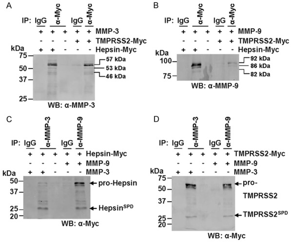
Figure 6.

MMP-3 and MMP-9 immunoprecipitate with hepsin and TMPRSS2. Western blot analyses were performed on cell surface biotinylated fractions from COS-7 cells transiently expressing proteases of interest. A. Lysates from COS-7 cells transiently transfected with constructs encoding MMP-3 and hepsin-MYC or TMPRSS2-Myc were subjected to immunoprecipitations using anti-Myc or control immunoglobulins (IgG) then examined by anti-MMP-3 Western blot analysis. ProMMP-3, 57 kDa; intermediate MMP-3, 53 kDa; activated MMP-3, 46 kDa. B. Lysates from COS-7 cells transiently transfected with constructs encoding MMP-9 and hepsin-MYC or TMPRSS2-Myc were subjected to immunoprecipitations using anti-Myc or control immunoglobulins (IgG) then examined by anti-MMP-9 Western blot analysis. Pro-MMP-9, 92 kDa; intermediate MMP-9, 86 kDa; activated MMP-9, 82 kDa. C. Lysates from COS-7 cells transiently transfected with constructs encoding hepsin-MYC and MMP-3 or MMP-9 were subjected to immunoprecipitations using anti-MMP-3, anti-MMP-9 or control immunoglobulins (IgG) then examined by anti-Myc Western blot analysis. D. Lysates from COS-7 cells transiently transfected with constructs encoding TMPRSS2-MYC and MMP-3 or MMP-9 were subjected to immunoprecipitations using anti-MMP-3, anti-MMP-9 or control immunoglobulins (IgG) then examined by anti-Myc Western blot analysis.
