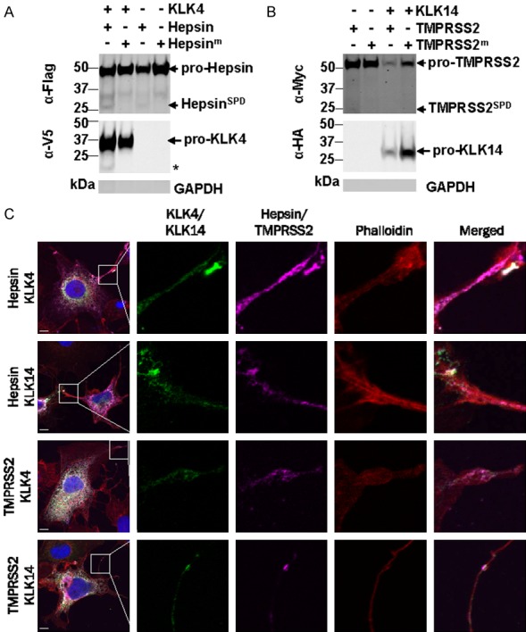
Figure 7.

Hepsin, TMPRSS2, KLK4 and KLK14 localize to the cell surface. A. Western blot analysis of cell surface biotinylated fractions from COS-7 cells transiently transfected with KLK4-V5 and hepsin-Flag. Asterisk, 17 kDa KLK4 cleavage products. B. Western blot analysis of cell surface biotinylated fractions from COS-7 cells transiently transfected with KLK14-HA and TMPRSS2-Myc. Purified fractions were free of GAPDH indicating that cells were intact during biotinylation. C. Confocal microscopy analysis of COS-7 cells co-expressing hepsin-Flag or TMPRSS2-Myc (purple) with KLK4-V5 or KLK14-HA (green). Cells were co-stained with DAPI to delineate cell nuclei (blue), and Alexa Fluor 568 conjugated phalloidin to delineate F-actin positive cytoplasm (red). White, regions of co-localisation of hepsin/TMPRSS2 (purple) with KLK4/KLK14 (green). Scale bar = 10 µm.
