Fig. 5.
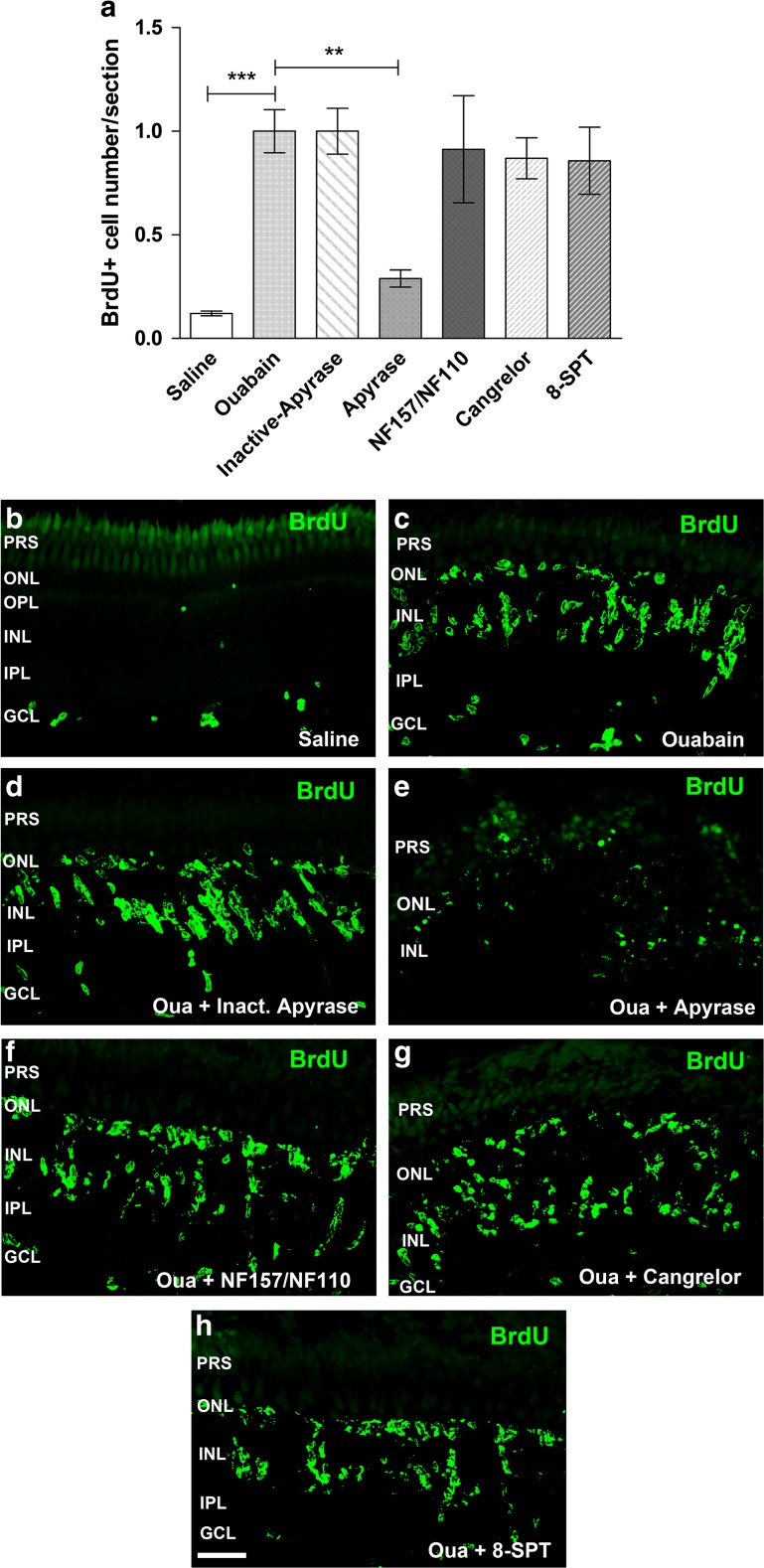
Apyrase and purinergic receptor antagonist treatment effect on in vivo cell proliferation after injury. Eyes underwent a single intravitreal injection of saline solution or ouabain on day 0. Eyes were injected daily for 6 days after lesion (dpl) with saline solution, apyrase (di- and tri-phosphate nucleotidase), heat-inactivated apyrase, or different purinergic receptor antagonists: cangrelor (P2RY12, P2RY13), 8-SPT (adenosine P1R), or NF110 plus NF157 (P2RX1, P2RX2, P2RX3, and P2RY11). A single dose of 5-bromo-2′-deoxyuridine (BrdU) was injected for 3 days starting on day 4 after injury together with the enzyme apyrase or the antagonists. Zebrafish were euthanized 7 days after injury. a Normalized number of BrdU-positive nuclei per retinal section. b–h Representative confocal images (bi-dimensionally reconstructed from z-stacks) of BrdU-labelled nuclei in 16-μm sections obtained from ouabain-treated retinas that were in vivo exposed to the different antagonists as indicated in each panel. Normalized data were expressed as mean ± SE (n = 4–8 zebrafish per group). ***p < 0.001, **p < 0.01, Dunnett’s multiple comparison test (vs. the ouabain treatment) after ANOVA. Scale bar 40 μm. PRS photoreceptor segments, ONL outer nuclear layer, OPL outer plexiform layer, INL inner nuclear layer, IPL inner plexiform layer, GCL ganglion cell layer, Oua ouabain

