Figure 2.

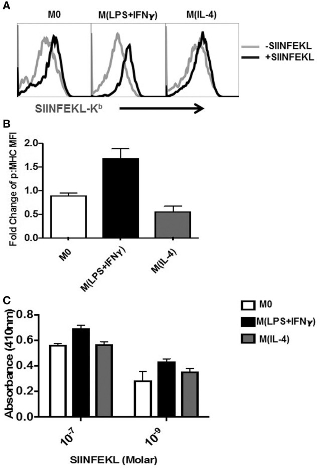
Detection of SIINFEKL peptide bound to MHC-I on MΦ. Sp-MΦ were polarized into either M(LPS + IFN-γ), M(IL-4) or left untreated (M0) and pulsed with SIINFEKL (10−7M) for 2 h at 37°C. (A) Cells were stained with 25-D1.16 monoclonal antibody, which detects SIINFEKL bound to H2-Kb MHC-I (p:MHC) before acquisition using FCM. The data are demonstrative histograms from one of three representative experiments. (B) Fold change in MFI of detected ab staining was calculated by comparing 25D staining in SIINFEKL pulsed versus unpulsed controls. Graphical data show mean ± SD from three independent experiments. (C) Cells were pulsed 10−7 or 10−9 M SIINFEKL for 2 h at 37°C before coincubation with the T-cell B3Z hybridoma for 18 h (1:1 ratio). The detection assay was carried out as described in Section “Materials and Methods” and OD was measured at 415 nm. Graphs depicting mean ± SD from three experimental replicates. MFI, mean fluorescent intensity; MHC, major histocompatibility complex.
