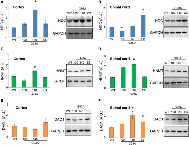
Figure 8.
Protein expression levels of histidine decarboxylase (HDC), histamine N-methyltransferase (HNMT), and diamine oxidase (DAO) in cortex and lumbar spinal cord from wild-type (WT) and SOD1-G93A mice. Cortex (A,C,E) and lumbar spinal cord (B,D,F) from WT (~20 weeks) and SOD1-G93A mice at different stages of the disease (100, 140 days, and end stage) (n = 4/group) were subjected to SDS-PAGE, western blotting, and immunoreactions with anti-HDC, anti-HNMT, and anti-DAO. Anti-GAPDH was used for protein normalization. Data represent mean ± SEM. Statistical significance was calculated by ANOVA followed by Post Hoc Tukey’s test, as referred to WT mice, *p < 0.05.

