
Fig. 4.
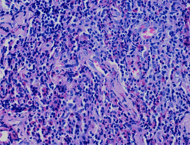
Interfollicular areas showing high vascularity with high endothelial venules and focal hyalinization. The inflammatory infiltrate is comprised predominantly of lymphocytes and many admixed eosinophils. (H&E stain, ×400)
Official websites use .gov
A
.gov website belongs to an official
government organization in the United States.
Secure .gov websites use HTTPS
A lock (
) or https:// means you've safely
connected to the .gov website. Share sensitive
information only on official, secure websites.
Interfollicular areas showing high vascularity with high endothelial venules and focal hyalinization. The inflammatory infiltrate is comprised predominantly of lymphocytes and many admixed eosinophils. (H&E stain, ×400)