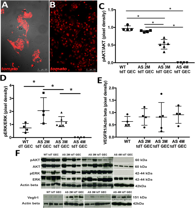
Figure 3.
VEGF signaling is altered in GEC isolated from Alport mice. VEGF signaling changes in GEC were studied using tdTomato-labeled GEC (tdT GEC) isolated by FACS from Alport-TektdT glomeruli (A, 10x). tdT signal (red) is strongly present in all cells as shown in (B) (10x, two passages in culture) (n = 3). Graphs representing immunoblot data of pAKT/AKT (C, 60 kDa/60 kDa), pERK/ERK (D, 42 kDa/42 kDa), VEGFR1 expression (E, 151 kDa) in glomeruli of tdT GEC derived from WT mice and Alport-TektdT mice at different ages. Imbalanced VEGF signaling is evident during disease progression in GEC, identified by a strong alteration in pAKT/AKT and pERK/ERK signaling downstream of VEGFR2. (F) Immunoblots of all the experimental groups used to generate the graphs presented in Figures C–E. Immunoblots were quantified by densitometry (VEGFR1 measurements were normalized against corresponding housekeeping gene, β-actin, 42 kDa). These data were obtained using GEC derived from n = 4 WT at 5 months of age, n = 4 AS mice at 2 months of age, n = 6 AS mice at 3 month of age, n = 4 AS mice at 6 months of age. One-way ANOVA with Tukey’s post hoc test was used to analyze the data and scatter plot values are presented as mean ± SD (*p < 0.05).

