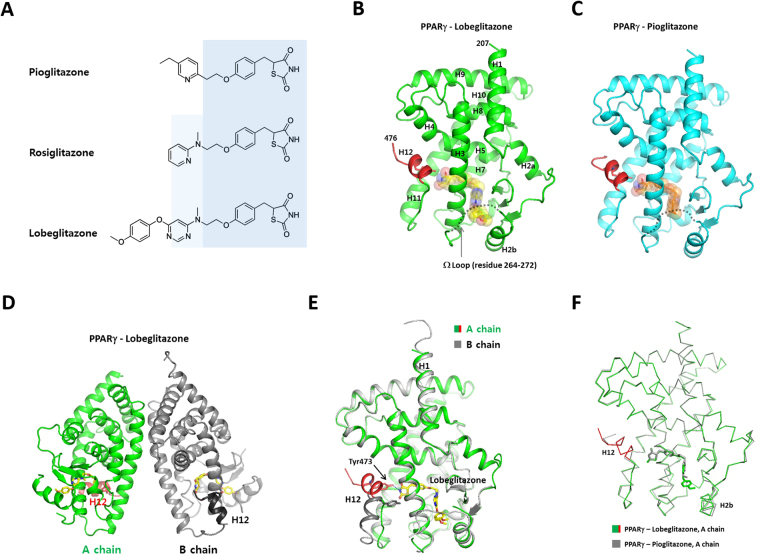
Figure 1.
Overall structures of the PPARγ LBD complexed with pioglitazone and lobeglitazone. (A) Chemical structures of pioglitazone, rosiglitazone, and lobeglitazone. Structurally conserved parts of TZDs are shaded with light blue colors. (B) Monomeric structure of the PPARγ LBDs complexed with lobeglitazone. The C-terminal AF-2 helix (H12) is colored in red. The disordered Ω-loops are indicated with dashed lines. The bound ligands are shown as transparent spheres. (C) Monomeric structure of the PPARγ LBDs complexed with pioglitazone. (D) The PPARγ LBD crystallized as a homo-dimer composed of active and inactive forms in an asymmetric unit. (E) Structural superposition of A and B chains of the lobeglitazone-bound PPARγ LBD. (F) Structural comparison of the lobeglitazone- and pioglitazone-bound PPARγ LBDs.

