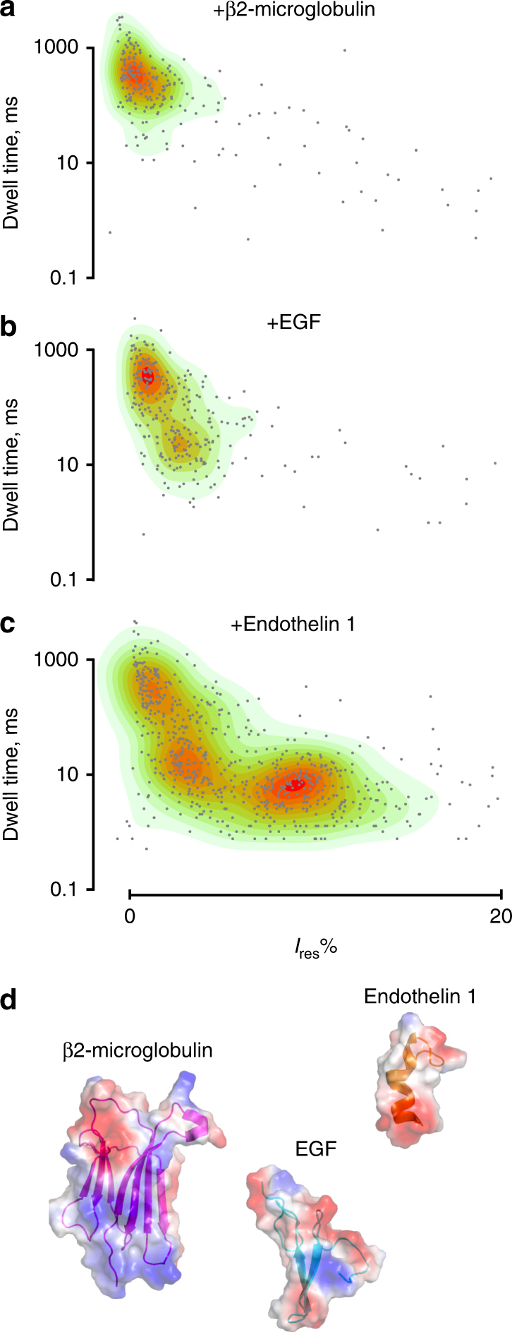
Fig. 4.

Discrimination of a biomarker mixture with WtFraC at pH 4.5. A single WtFraC nanopore was obtained in a buffer at pH 4.5 (1 M KCl, 0.1 M citric acid, 180 mM Tris base) under a −50 mV applied potential. About 200 nM of β2-microglobulin was initially added to the cis compartment (a), then 1 µM EGF (b), and finally 200 nM endothelin 1 (c) were added to cis compartment. d Crystal structure of β2-microglobulin, EGF, and endothelin 1 mixture created with PyMOL colored according to their vacuum electrostatics
