Figure 1.
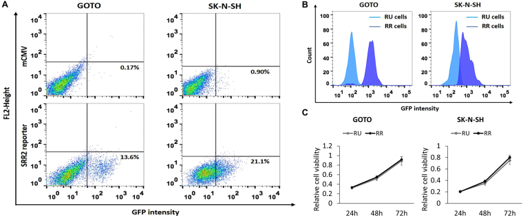
Identification of two cell subsets in NB cell lines. (A) Flow cytometry analysis was performed to detect GFP expression in NB cells stably infected with the lentiviral SRR2 reporter. In both cell lines, a small subset of reporter responsive (RR) cells was found, whereas the majority of the cells were unresponsive to the reporter (i.e. reporter unresponsive, RU cells). NB cells stably infected with the mCMV (negative control) lentivirus were used as the negative control. (B) GFP expression in purified RU and RR cells purified from cells stably infected with the lentiviral SRR2 reporter were substantially different. (C) Cell growth of RU and RR cells were assessed by using the MTS assay. All data are presented as mean ± SD, *p < 0.05, Student’s t test. Abbreviations: NB, neuroblastoma; SRR2, Sox2 regulatory region 2; mCMV: Murine Cytomegalovirus; GFP: Green Fluorescence Protein.

