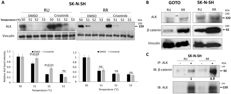
Figure 3.
RR cells demonstrate no crizotinib—ALK binding and higher expression level of β-catenin compared to RU cells. (A) CETSA was performed to compare crizotinib—ALK binding ability between RU and RR cells. RU and RR cells derived from SK-N-SH were treated with DMSO or 500 nM crizotinib for 6 hours. Representative ALK western blots are shown on the upper panel. Vinculin level was blotted as a loading control. The densitometry quantification data from 3 independent experiments are shown on the lower panel. All data are presented as mean ± SD. Student’s t test was performed. (B) The expression of ALK and β-catenin protein in RU and RR cells were measured by western blots. Vinculin level was blotted as a loading control. (C) ALK pull-down was performed using co-immunoprecipitation assay and showed substantial ALK-β-catenin binding in RR cells compared to RU cells.

