
Figure 6.
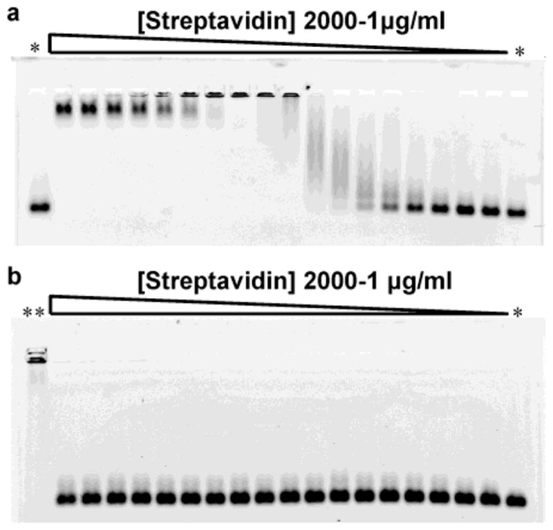
Streptavidin gel retardation assay with biotinylated peptide-coated nanocrystals. (a) 1% agarose gel electrophoresis of biotinylated red emitting CdSe/ZnS nanocrystals coated with peptide 8. The incubation of the nanocrystals with increasing concentration of streptavidin leads to the formation of aggregates and a shift of the nanoparticle band. Large nanocrystal aggregates are unable to enter the gel pores. The gel re-entry at high streptavidin concentration corresponds to the saturation of the nanocrystal surface biotins that prevents aggregation. (b) 1% agarose electrophoresis of nonbiotinylated CdSe/ZnS nanocrystals coated with peptide 6 under the same condition as in (a). The concentrations of streptavidin tested were 2000, 1000, 750, 500, 300, 250, 150, 100, 50, 40, 30, 25, 20, 15, 10, 5, 2 and 1 μg/mL. *: no streptavidin; **: biotinylated nanocrystals + 250 μg/mL streptavidin.