Figure 3.

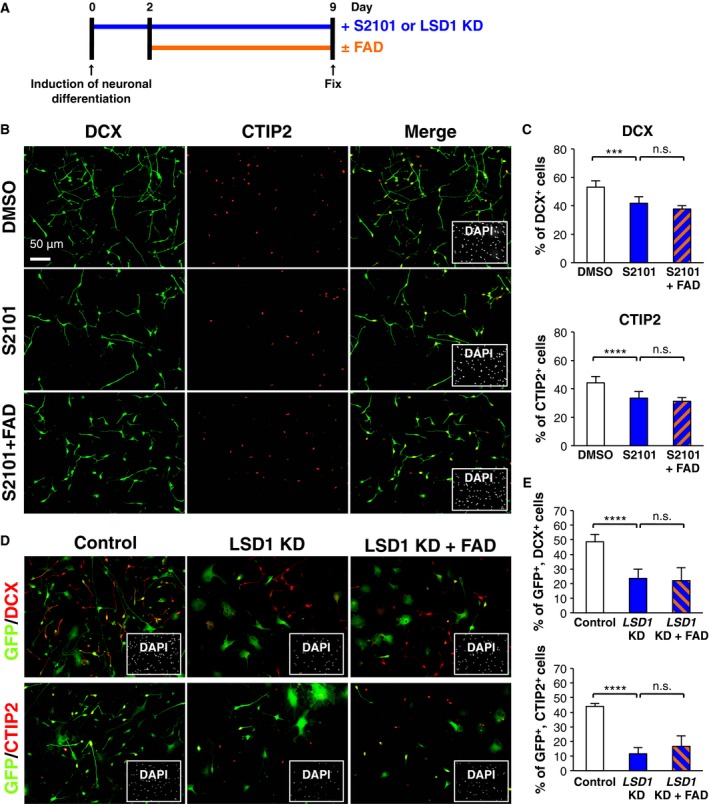
Promotion of neuronal differentiation by FAD administration mediates LSD1 activity. (A) Diagram depicting the cell culture and neuronal differentiation schedule. (B) The cells treated with S2101, FAD, or their combination were stained with antibodies against DCX (green) and CTIP2 (red) at 9 days after induction of differentiation. Insets: Hoechst nuclear staining of each field. Scale bar: 50 μm. (C) The graphs indicate the percentage of total cells that were DCX‐ and CTIP2‐positive. The values shown are the means ± SD. (N = 6, ANOVA, ***P < 0.005, ****P < 0.001, n.s.: not significant). (D,E) The cells, infected with recombinant lentiviruses engineered to express only GFP or GFP with shRNA designed against LSD1, were stained with antibodies against GFP (green) and DCX (red in upper panel) or CTIP2 (red in lower panel) at 7 days after induction of differentiation. The percentage of GFP‐positive cells that were DCX‐ or CTIP2‐positive was calculated. The values represent the means ± SD. (N = 6, ANOVA, ****P < 0.001, n.s., not significant).
