
Figure 3.
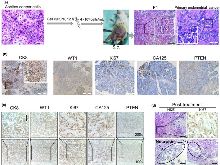
Ascites‐derived tumor cells are tumorigenic and patient‐derived xenografts mirror patient response to therapy in a 62‐year‐old woman with endometrial cancer and breast cancer. (a) Ascites‐derived tumor cells enriched from the patient were injected into NOD/SCID mice. Representative images of H&E staining are shown. (b,c) Representative images of immunohistochemistry staining in primary endometrial cancer (b) and the patient‐derived xenograft model (c). CA125, cancer antigen 125; CK8, cytokeratin 8; PTEN, phosphatase and tensin homolog; WT1, Wilms’ tumor suppressor gene 1. (d) Prominent central necrosis in the treated group and immunohistochemistry staining of anti‐Ki67.