
Figure 6.
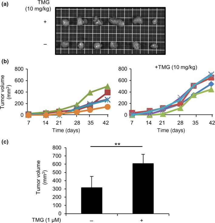
Transplanted LoVo cell‐derived tumor formation was accelerated in Thiamet G (TMG)‐treated BALB/c‐nu/nu mice. LoVo cells were s.c. transplanted onto the backs of Balb/c‐nu/nu mice (n = 6), with or without daily peritoneal TMG treatment, under anesthesia with tribromoethanol (300–400 mg/kg), i.p. (a) Tumors derived from LoVo cells were s.c. transplanted into BALB/c‐nu/nu mice. Tumors (n = 6, each group) were then surgically removed from mice treated with or without TMG. Images of the removed tumors are shown (top row, TMG treatment; bottom row, control). (b) The size of each tumor was measured weekly starting 1 week after transplantation. Tumor size was calculated using the formula (width × depth2/2; width > depth). (c) Average tumor volume (n = 5) was calculated. Tumors with sizes that deviated from the mean ± SD were excluded in each group. **P < 0.01, Student's t‐test.