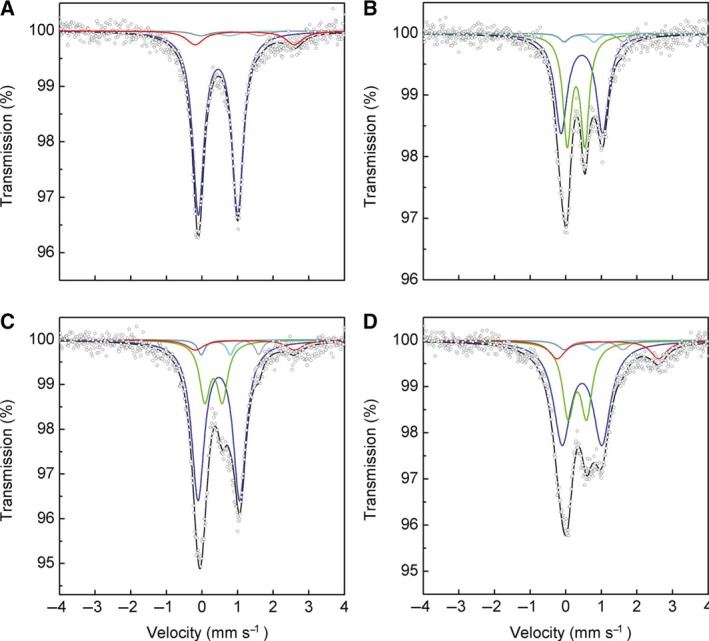
Figure 1.

Mössbauer spectra of WT and C32S mutant of PqqE. 57Fe Mössbauer spectra of (A) 56Fe‐as‐purified WT enzyme reconstituted with 57Fe, (B) 57Fe‐as‐purified WT enzyme reconstituted with 56Fe, (C) 57Fe‐as‐purified WT enzyme reconstituted with 57Fe, and (D) 57Fe‐as‐purified C32S mutant reconstituted with 57Fe, all recorded at 5 K. Simulated spectra are shown as follows: blue, [4Fe–4S]2+; green, [2Fe–2S]2+; gray and light blue, ferrous and ferric sites, respectively, of the [4Fe–4S]2+ cluster ligated with the side‐chain carboxyl group of Asp319 at the Aux II site; red, free (noncluster) high‐spin Fe2+; black, the sum of the simulated spectra.
