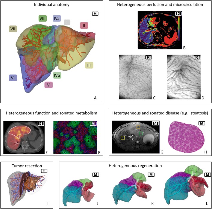
Figure 2.
Spatial heterogeneity in liver physiology. Visualization of human individual hepatic vascular and parenchymal anatomy (A, the labels indicate the different Couinaud segments) is the basis of current surgical planning (I). Planning currently does not take any functional heterogeneity into account. However, heterogeneity exists on the macro- and microscale in terms of hepatic perfusion (B, clinical perfusion CT*) and microcirculation [C,D, orthogonal polarization spectroscopy image from (C) normal rat liver and (D) rat liver after 90%PHx]. Heterogeneity also occurs in terms of regional distribution of functional activity (E, Mebrofenin scan of human liver**) and of metabolic zonation in mouse liver (F, periportal expression of E-cadherin and perivenous expression of CYP2E1). Furthermore, inhomogeneous distribution also occurs in case of morphologic changes due to global liver disease, here shown regional heterogeneity of fat distribution (G, MRT of steatotic mouse liver) as well as zonated distribution of fat accumulation in periportal hepatocytes in a mouse liver (H). Current planning focuses on visualizing tumor location (I). Monitoring of liver regeneration is mostly restricted to experimental or clinical studies and revealed inhomogeneous growth of the remnant lobes in mice (J–L). H, human; M, mouse; R, rat. *Reprinted from Cieslak et al. (2016), with permission from Elsevier. **Reprinted from Wang et al. (2013), with permission from Elsevier.

