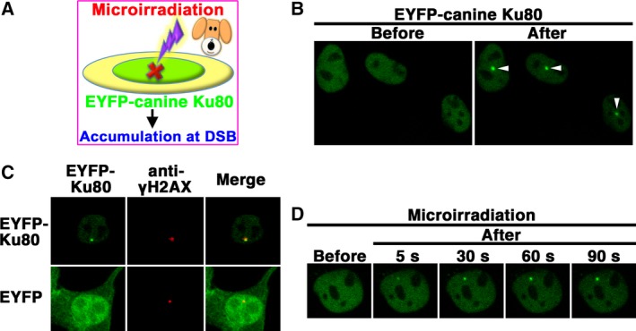
Figure 4.

Accumulation of EYFP‐canine Ku80 at the sites of DSBs induced by laser microirradiation. (A) The recruitment of EYFP‐canine Ku80 to DSBs induced by 405‐nm laser irradiation in MDCK cells. (B) Imaging of live MDCK cells transfected with pEYFP‐canine Ku80 before (left panel) and after (right panel) microirradiation. Arrowheads indicate the microirradiated sites. (C) Immunostaining of microirradiated cells transfected with pEYFP‐canine Ku80 using an anti‐γH2AX antibody. Cells were fixed and stained with an anti‐γH2AX antibody 5 min postirradiation. Left panel, EYFP‐canine Ku80 (upper panel) or EYFP (lower panel); center panel, γH2AX; right panel, merged images. (D) Time‐dependent EYFP‐canine Ku80 accumulation in live cells, from 5 to 90 s after irradiation.
