Figure 1.

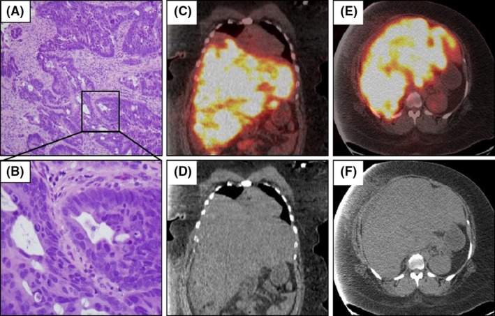
Biopsy of colonic mass and fused PET/CT images. H&E staining of colon mass at lower (A) and 40× (B) magnification. Fused PET‐CT sagittal (B,C) and coronal sections (D,E) revealing very avid uptake in the liver.

Biopsy of colonic mass and fused PET/CT images. H&E staining of colon mass at lower (A) and 40× (B) magnification. Fused PET‐CT sagittal (B,C) and coronal sections (D,E) revealing very avid uptake in the liver.