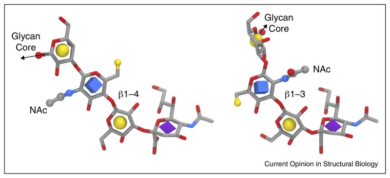
Figure 3.
HA receptor structures indicating the influence of the Gal-2 — GlcNAc3 linkage type (left: β1-4, right: β1-3) on conformation and presentation. The structures were retrieved from PDB IDs 4YYA and 4NRL, respectively, and aligned relative to the Sia residues. Note the reversal of the N-acetyl moieties relative to the Sia residues. The GlcNAc 6-position, which may be sulfated, is shown as a small yellow sphere.

