Fig. 2.

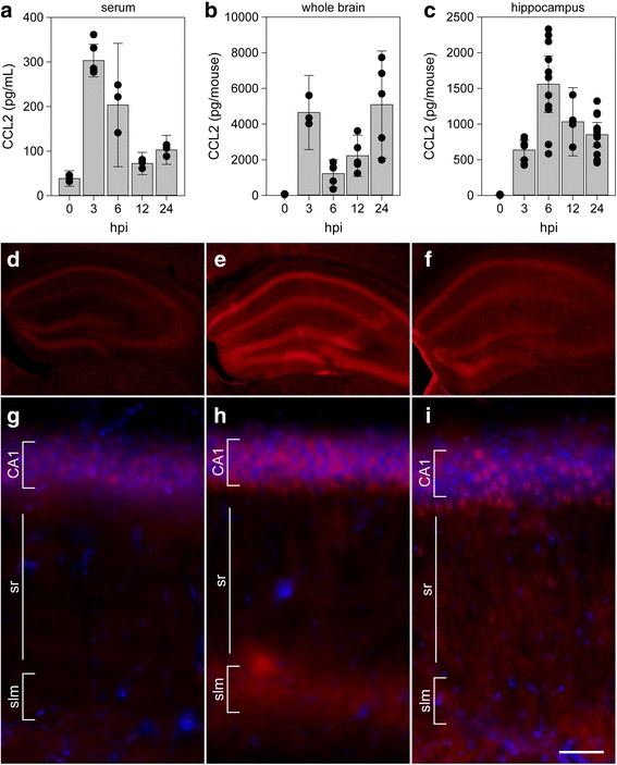
CCL2 levels in serum, whole brain, and hippocampus during acute infection. Serum (a), whole brain (b), and microdissected hippocampus (c) were collected at 0, 3, 6, 12, and 24 h following intracranial inoculation with TMEV. Absolute levels of CCL2 were measured by ELISA. Each dot represents an individual animal. The bar graph shows mean ± 95% confidence intervals from aggregate data. A minimum of three mice were used per tissue per timepoint, and the hippocampal samples were collected in a separate experiment from the whole brain samples. All results were analyzed by one-way ANOVA. CCL2 was induced in serum, whole brain, and hippocampus (P < 0.001 for each factor in each tissue compared to 0 hpi). Brain sections collected at 0 (d, g), 6 (e, h), and 24 hpi (f, i) were immunostained to reveal CCL2 expression. In the hippocampus, there was weak, diffuse signal in uninfected mice (d, g) that increased markedly at 6 hpi (e, h). By 24 hpi, the signal intensity had decreased and exhibited a more punctate pattern (f, i). Scale bar in i is 50 μm and refers to g–i. CA1 cornu ammonis 1 formation, sr stratum radiatum, slm stratum lacunosum moleculare. Immunostaining is representative of more than three mice per timepoint in more than three separate experiments
