Figure 6.
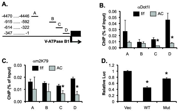
Dot1l represses Atp6v1b1 at least in part by modulating targeted H3 K79 hypermethylation at the Atp6v1b1 promoter. (A) Diagram of the 5′ flanking region of Atp6v1b1 that encodes V-ATPase B1. (B & C) ChIP demonstrating impaired binding of Dot1l and H3m2K79 in the Atp6v1b1 5′ regulatory region. Chromatin from adult Dot1lf/f and Dot1lAC mice (n=6 mice/group) was immunoprecipitated by the rabbit antibodies specific for Dot1l (B) and H3m2K79 (C), followed by real-time qPCR with primers amplifying subregions A–D as shown in A. Relative ChIP efficiency was defined as the immunoprecipitated amount of materials present as compared to that of the initial input sample. *: P<0.05 vs. Dot1lf/f within the same subregion. (D) Luciferase assay showing that Dot1a represses a luciferase construct driven by a 0.84kb-promoter of Atp6v1b1 in IMCD3 cells. IMCD3 cells stably carrying the luciferase construct were transfected with an empty vector, or a construct expressing WT or methyltransferase-dead mutant Dot1a (MT) and analyzed for luciferase activity (n=3). *: P<0.05 vs. Vec.

