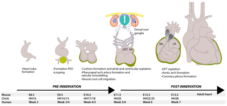
Figure 1.
Development of the heart and neural tube in mice. At E8.5, the murine heart is a tube with blood flowing in a peristaltic manner from the caudal venous pole to the cranial arterial (outflow) tract. At E9.5, the heart starts looping and, at the same time, epicardial cells from the proepicardial organ (green) start to migrate and cover the outside of the heart. At E11.5, neural crest-derived cells (blue) delaminate from the neural tube and start migrating ventrally and caudally, contributing to many structures, including dorsal root ganglia. They contribute to valvular remodeling and the septation of the pulmonary and aortic vessels, as well as to delivering neurons to innervate the heart. When the heart has finished looping, the inflow and outflow tract are both found at the base of the heart and the electrical conduction now has an apex-to-base direction. DRG = dorsal root ganglion, HH = Hamburger and Hamilton stage, LA = left atrium, LV = left ventricle, OFT = outflow tract, RA = right atrium, RV = right ventricle, PEO = proepicardial organ.

