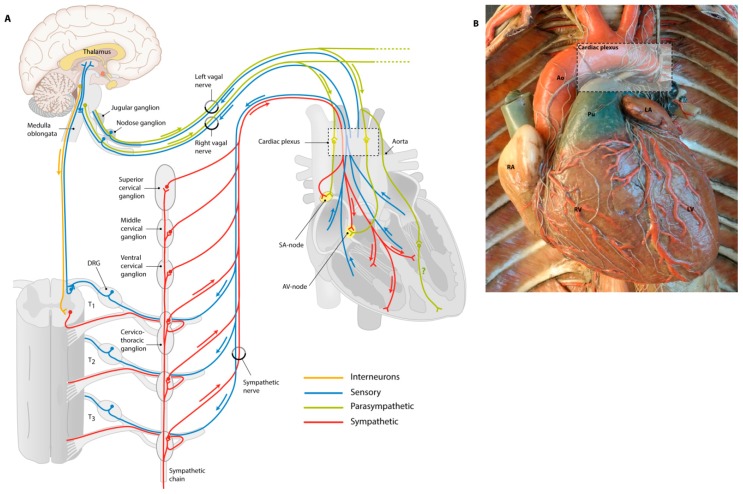
Figure 2.
Overview of cardiac innervation. (A) Schematic drawing of the cardiac visceral innervation system. Cardiac innervation starts with a signal from the heart or baroreceptors (e.g., on the aorta), relayed by sensory nerves (blue) giving feedback on, for instance, the levels of oxygen, carbon dioxide and blood pressure. The brain will give a signal to parasympathetic or sympathetic nerves to either relax or stimulate the heart. Parasympathetic innervation is achieved mainly via the vagal nerve (green) that will synapse in cardiac ganglia from where postganglionic nerves innervate the SA node and AV node, and potentially ventricular myocytes. Sympathetic neurons (red) start in the grey matter of the spinal cord, where interneurons (orange) from the brain project to the sympathetic neurons. Via the ventral root of the spinal cord, sympathetic nerves synapse in the sympathetic chain, from where postganglionic nerves will enter the heart; (B) This wax mold shows how cardiac nerves enter via the cardiac plexus and follow cardiac vessels over the heart. Courtesy of: Museo delle Cere Anatomiche “Luigi Cattaneo”, University Museum System, Alma Mater Studiorum—University of Bologna, picture taken by Dr. E.A.J.F. Lakke. Ao = aorta, DRG = dorsal root ganglion, LV = left ventricle, Pu = pulmonary artery, RA = right atrium, RV = right ventricle.

