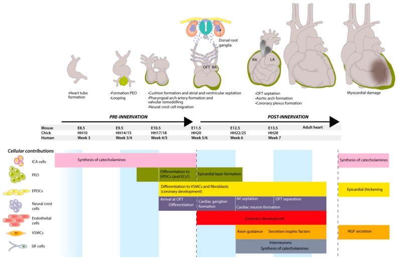
Figure 8.
A summary of the development of cardiac innervation and cellular contributions. At every developmental stage, different cell populations are involved. Before cardiac innervation is established, intrinsic cardiac adrenergic (ICA) cells already contribute to the autonomic regulation by synthesizing catecholamines. Additionally, after transplantation, these cells become active again and produce tyrosine hydroxylase (TH) and dopamine-β-hydroxylase (DBH). The proepicardial organ (PEO) is important for the coronary development, but also the source of epicardial-derived cells (EPDCs) and maybe endothelial cells (ECs). The formation of the epicardial layer is a hallmark for cardiac innervation. EPDCs are a major source of several cells, including vascular smooth muscle cells (VSMCs) and fibroblasts, thereby also contributing to the coronary development. Neural crest cells play a multi-facetted role in the development of the heart and cardiac innervation. VSMCs are mainly important for axon guidance through secretion of neurotrophic factors, but are also a player in pathological hyperinnervation by secretion of nerve growth factor (NGF) after myocardial damage. Small intensely fluorescent (SIF) cells can act as interneurons and are together with ICA cells a cardiac source for catecholamines. DRG = dorsal root ganglion, OFT = outflow tract.

