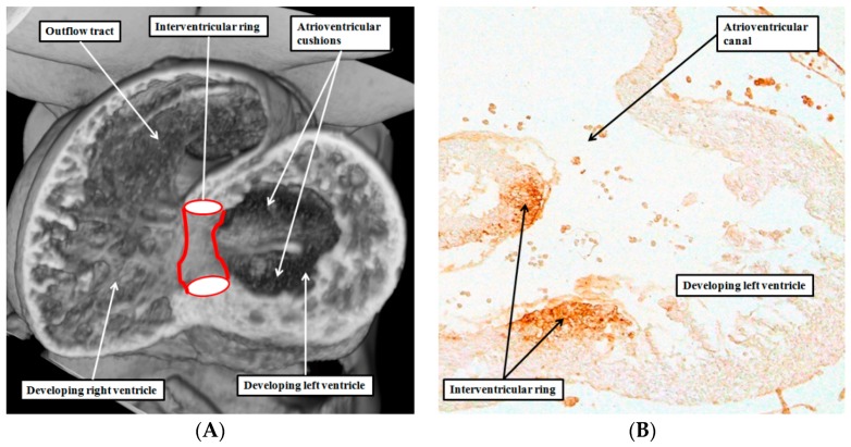
Figure 1.
The left hand panel (A) is taken from an episcopic dataset prepared from a mouse embryo at E10.5. It is cut along the long axis of the ventricular loop, and shows the location of the interventricular ring, which has the inner heart curvature as its cranial margin, and the developing muscular ventricular septum as the caudal margin; The right hand panel (B) is a comparable section from a human embryo processed with an antibody to the nodose ganglion of the chick heart. It shows that the cranial margin of the interventricular ring not only occupies the inner heart curvature, but also forms the rightward margin of the atrioventricular canal. The original section was prepared in Amsterdam by Professor Andy Wessels, and is reproduced with his permission.

