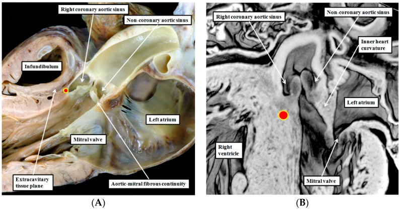
Figure 4.
The left hand panel (A) shows the outflow tract of the normal left ventricle sectioned to replicate the so-called parasternal long axis echocardiographic plane. It shows the fibrous continuity between the aortic leaflet of the mitral valve and the non-coronary leaflet of the aortic valve. The red circle with yellow borders shows the location of the dead-end tract. Note the extracavitary tissue plane between the crest of the ventricular septum and the subpulmonary infundibulum; The right hand panel (B) is taken from an episcopic dataset prepared from a developing mouse embryo at E15.5. By this stage, the embryonic interventricular communication is closed, but the inner heart curvature forming the roof of the left ventricle, and interposing between the developing leaflets of the aortic and mitral valves, remains myocardial. The red circle with yellow borders again shows the location of the dead-end tract.

