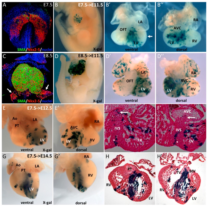
Figure 1.
Early SMA+ cardiomyocytes contribute to the interventricular septum. (A–C) Whole-mount immunofluorescence reveals early expression of SMA in Nkx2-5+ cells. At E7.5, SMA is expressed in a subpopulation of cells in the cardiac crescent and, at E8.5, in the early heart tube with the exception of the venous pole (arrows). (B–D) Whole-mount X-gal staining of E11.5 SmaCre/+::R26RLacZ/+ embryos after tamoxifen injection at E7.5 (B) or E8.5 (D). (B’,B’’) ventral and dorsal views of an E11.5 heart showing X-gal labeling in the left ventricle (LV) and interventricular septum (IVS). No labeling was observed in left ventricular free wall myocardium (arrow). (D’,D’’) ventral and dorsal view of E11.5 heart with X-gal labeling throughout the heart with the exception of the OFT. Ventral (E,F) and dorsal (E’,F’) whole-mount and serial sections of E12.5 SmaCre/+::R26RLacZ/+ embryos after tamoxifen injection at E7.5. X-gal labeled cells are present in the LV and IVS with sparse positive cells at the upper part of the septum (arrows), in contrast to compact vertical clusters at the base of the IVS (stars). Ventral (G,H) and dorsal (G’,H’) whole-mount and serial sections of an E14.5 SmaCre/+::R26RLacZ/+ embryo after tamoxifen injection at E7.5. X-gal labeling is intense in the IVS and few clusters are observed in the LV.

