Figure 3.
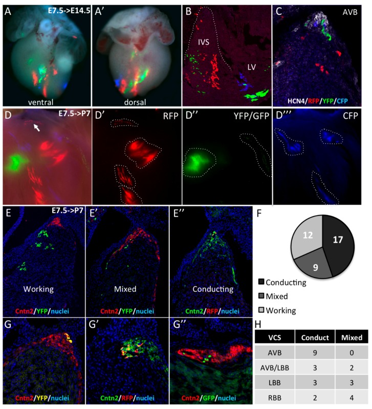
Early segregation of the central ventricular conduction system lineage in SMA+ cardiomyocytes. (A,A’) Whole-mount ventral and dorsal views of an E14.5 SmaCre/+::R26Rconfetti/+ heart with multicolor clusters induced by tamoxifen injection at E7.5. The four fluorescent reporters (GFP, YFP, RFP, and CFP) form independent clusters distributed side by side along the vertical axis in the apical interventricular region. (B) Section of an E14.5 SmaCre/+::R26Rconfetti/+ embryonic heart showing large vertical compact multicolor clones in the apical region of the IVS and scattered labeled cells in the upper part of the septum. (C) Hcn4 immunofluorescence on section of E14.5 SmaCre/+::R26Rconfetti/+ heart showing a heterogeneous contribution of labeled cells to the AVB and BB. (D) Example of clonal labeling in a P7 SmaCre/+::R26Rconfetti/+ heart with a small number of labeled clusters of which one RFP+ cluster is localized at the crest of the septum (arrow). Individual fluorescent reporters are presented in D’ (RFP), D’’ (GFP/YFP) and D’’’ (CFP). (E,G) Cntn2 immunofluorescence on sections from P7 SmaCre/+::R26Rconfetti/+ hearts to classify the nature these clusters. All cells in working clusters are negative for Cntn2 (E), few are positive in mixed clusters (E’) and all are positive for Cntn2 in conducting clusters (E’’). The distribution of these clusters is presented in the graph in the right panel (F). (G,G’,G”) Sections through independent conducting clones of different colors (YFP in G; RFP in G’ or GFP in G’’) showing co-labeling with Cntn2 in the AVB. (H) Table showing the distribution of mixed and conducting clones in different compartments of the central VCS.

